The Scary Stuff
Late on the night of March 23, I was laying in a hospital bed waiting the results of a brain MRI. The day had been so far outside my normal experience that to call it "extraordinary" borders on trivialization. That morning I walked into the Emergency Room looking for an explanation for bizarre symptoms that were progressing rapidly. My doctors had ordered a brain MRI, to rule out "the scary stuff," but it wasn't going to happen for weeks. I wasn't sure I had that kind of time. I'd blown past the triage nurse and right into a semi-private hospital room. A CT scan revealed something of enough concern to call for an immediate brain MRI.
Around 10:30 PM, a doctor I hadn't seen before entered the room holding a smart phone. On its screen was an image depicting a slice through my brain taken just minutes before. Even from the distance I was viewing, something was horribly, monstrously wrong. It occurred to me that this is the kind of thing a doctor thinks about when using the term "scary stuff" with a patient.
The white blob in the lower-left was described to me as a 5.1 cm long "legion" with dead tissue inside. It was roughly the size of a lemon. The word "cancerous" was used. There's no fucking way this is real.
On the one hand I couldn't believe what I was seeing or hearing. Normal brains don't look like that. Normal brains have bilateral symmetry. The left half looks like the mirror image of the right half. Normal brains don't have legions the size of a lemon stuck in them. Normal brains don't have large volumes of dead tissue buried inside them. How was I even alive with that thing inside?
On the other hand it was a relief to shine a spotlight on my attacker. What brought me to this point was so strange that I had started to wonder if I might be imagining all of the symptoms. The MRI proved that I was imagining nothing. The presence of the lesion on the right side of the brain even explained the unilateral left-side symptoms.
Interlude
From this point on, brain MRI images are going to play a very important role in the story. So now seems like a good time to push the pause button on the horror unfolding before me in that hospital bed on a late night in March 2023 for a quick backgrounder.
To the extent I had previously considered MRI before all of this, I thought of it as yielding a kind of 3D model that could be rotated and manipulated — a bit like the digital analog of those clear plastic anatomy models used in elementary schools. This is not generally correct. Instead, think of MRI as a way to virtually slice an object in two, as if with a very sharp and flat knife. An MRI image is what you'd see by separating the slices, then looking at the surface of the one facing you.
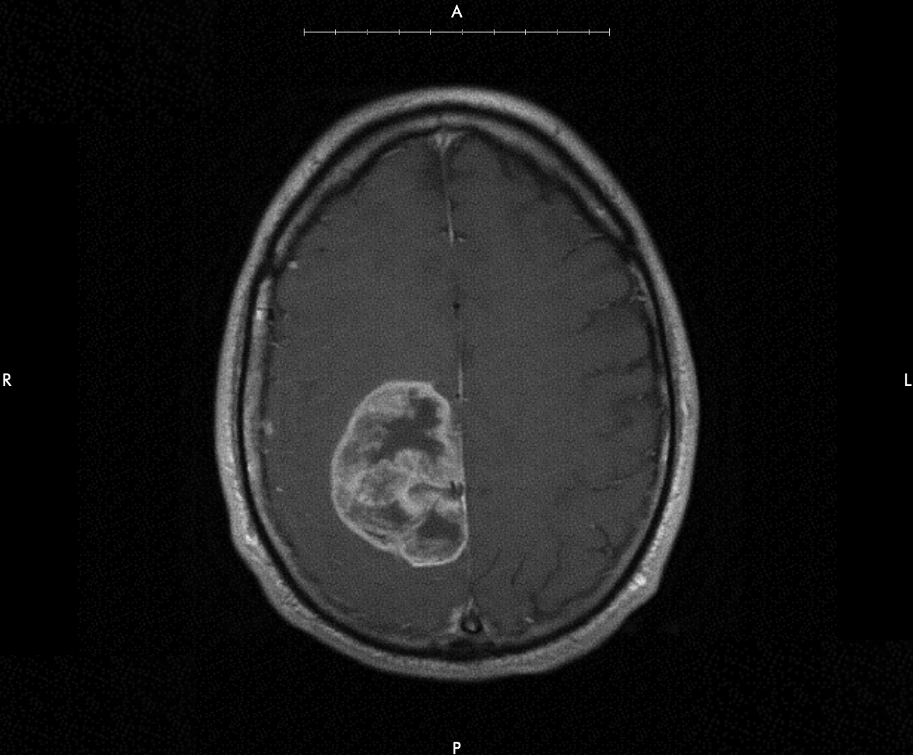
In a brain MRI the object being virtually sliced is a human brain; in this case, mine. Imagine I were laying on my back on a table in front of you. There would be several ways you might consider slicing through me. One useful way would be to slice perpendicular to the long axis of my body running from the soles of my feet to the top of my head. Making such a slice through my head would cut m my brain into two pieces, a bottom and a top. The name for this kind of slice is "axial," which is also the name of the view. An axial MRI shows the surface of the top brain slice.
Axial MRI views follow certain conventions, which among other advantages makes them easy to compare. The surface being shown is the one you'd get by looking from the soles of the feet to the top of the head with the patient lying on their back. The patient's right side is on your left, and vice versa. The nose points up, and the back of the head points down. You're then viewing the surface of the slice containing the top of the head. To avoid confusion, the borders of an axial brain MRI are labeled with the letters "R" (patient right); "L" (patient left); "A" (patient anterior); and "P" (patient posterior).
I'll have a lot more to say about brain MRIs in the posts to follow.
Taking Stock
This quick orientation makes the image I was seeing easier to understand. I had a large cancerous legion on the right side of my brain, near the back.
I entered the hospital that morning expecting to be sent home for rest and fluids at worst. Now I had brain cancer.